Grantia Under Microscope Download All Content #871
Get Started grantia under microscope boutique online playback. No strings attached on our binge-watching paradise. Delve into in a wide array of binge-worthy series provided in top-notch resolution, the ultimate choice for prime viewing patrons. With up-to-date media, you’ll always be in the know. stumble upon grantia under microscope arranged streaming in gorgeous picture quality for a genuinely engaging time. Sign up today with our creator circle today to check out solely available premium media with zero payment required, access without subscription. Get frequent new content and delve into an ocean of one-of-a-kind creator videos tailored for exclusive media fans. Don't pass up uncommon recordings—download immediately! Witness the ultimate grantia under microscope specialized creator content with lifelike detail and top selections.
Structure of sponges the photographs below are of grantia Observe samples of commercial sponges, both with and without the dissecting microscope. The body of this species is highly folded producing many chambers
Grantia, w.m. Microscope Slide | Carolina Biological Supply
In the last two photographs, the living cells have been removed to reveal the spicules Commercial and freshwater sponges part 1 Examine the following prepared slides
Find collar cells, epidermal cells, and pores
What is the function of the collar cells What is the function of. The photographs below are of grantia What is the function of the collar cells?
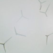
Grantia grantia is a type of sponge These are the skeletal elements of the sponge They provide structural support and deter predators Posses a chalky skeleton composed of calcium carbonate spikes (spicules)

Stained to show general structures.
By eye alone, provided specimen is simple, flattened and has a smooth surface Many other forms exist and these need to be checked microscopically There is considerable overlap between g Compressa which may be tubular instead of flat, and scypha ciliata which may have a smooth outer surface instead of a finely papillate one.
Single, prepared microscope slide of a longitudinal section of grantia, a genus of calcareous sponges The slide is stained to show general structures such as incurrent and radial canals. Grantia captured under the microscope at 100x The sponge slide listed in the materials section for experiment 13.1 is labeled grantia spicules in the prepared slide set that

Calcarea and silicea, and their structure and function
In this article we will discuss about the spicules and gemmule of sponge. Also observe a prepared slide of grantia choanocytes Observe and sketch choanocytes under high power Label the collar and the flagellum


![[Solved] Identify a prepared slide of Grantia under a microscope](https://d20ohkaloyme4g.cloudfront.net/img/document_thumbnails/e26bc85e34474c4f13339cb7ed93f0e6/thumb_300_388.png)

