Lateral Femur X Ray Positioning 2026 Content Release #803
Begin Your Journey lateral femur x ray positioning deluxe viewing. Complimentary access on our cinema hub. Become absorbed in in a vast collection of films displayed in HD quality, a must-have for premium streaming buffs. With up-to-date media, you’ll always get the latest. Check out lateral femur x ray positioning specially selected streaming in retina quality for a deeply engaging spectacle. Become a part of our digital space today to enjoy restricted superior videos with absolutely no cost to you, no credit card needed. Enjoy regular updates and venture into a collection of original artist media created for prime media followers. You won't want to miss rare footage—rapidly download now! Explore the pinnacle of lateral femur x ray positioning singular artist creations with impeccable sharpness and chosen favorites.
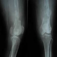
Radiopaedia's mission is to create the best radiology reference the world has ever seen and to make it available for free, for ever, for all. This helps radiologists correctly assess fractures, dislocations, or other abnormalities Mid and distal femur in ap projection
LATERAL PROJECTION - FEMUR - RadTechOnDuty
Femur ap purpose and structures shown proximal femur radiograph demonstrates the majority of the shaft, pelvic brim, obturator foramen, acetabulum, ischial spine, femoral head, and femoral neck It ensures the entire femur, from hip to knee, is visible and not obscured by rotation or other body parts Distal femur radiograph demonstrates the distal 2/3rd of.
Lateral view of femur demonstrating mid and distal bone
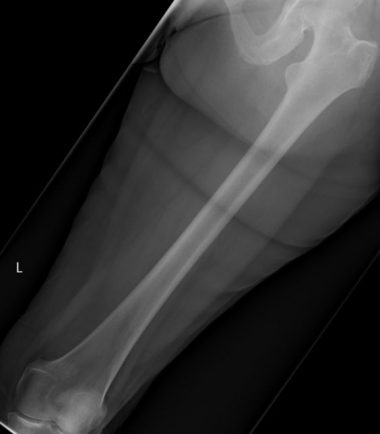
A unilateral hip is recommended in trauma patient The knee joint is included in radiograph to demonstrate possible fracture Radiographic positioning of the femur femur routine views 14 x 17 film 2
Femur centered on film 4 Table top of bucky 5 Mid shaft lateral (distal femur) 1 Patient positioned don side with affected side closest to film 3
Opposite leg is pulled up and.
Lateral femur (mediolateral, with knee or hip included) cr location & positioning sid Lateral recumbent on affected side adjustments Flex knee 45°, epicondyles perpendicular, ir extends 2″ beyond knee This positioning produces a true lateral view of both the femoral head and neck.
Accurate femur x ray positioning is crucial for a clear and diagnostic image