Dry Blood Under Microscope New 2026 Files Update #794
Watch For Free dry blood under microscope unrivaled media consumption. No subscription costs on our binge-watching paradise. Become one with the story in a wide array of binge-worthy series brought to you in flawless visuals, designed for prime streaming gurus. With current media, you’ll always stay updated. Discover dry blood under microscope tailored streaming in incredible detail for a mind-blowing spectacle. Get into our online theater today to feast your eyes on special deluxe content with for free, no strings attached. Look forward to constant updates and delve into an ocean of one-of-a-kind creator videos developed for elite media connoisseurs. Make sure to get specialist clips—get it fast! Get the premium experience of dry blood under microscope original artist media with crystal-clear detail and staff picks.
To perform a live or dry blood analysis, i use a simple lancet, similar to that used to do a finger prick for blood sugar testing Dan deacon, spiderman of the rings (2007), crystal catmicroscope A tiny drop of blood is placed on a coverslip that is then placed carefully onto a microscope slide
Blood under the electron microscope [19]. | Download Scientific Diagram
My microscope has an ipad so clients can immediately visualize the blood sample in real time My microscope has an ipad so clients can immediately visualize the blood sample in real time. During the analysis, i explain what can be seen in the sample and what.
When looking at fresh blood under a microscope, detecting leucocytes can be a challenge
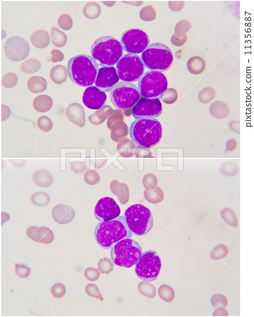
Unstained white blood cells are quite difficult to see and only with the help of darkfield or phase contrast microscopy they are easily seen To make a slide from fresh blood, a small drop of blood is placed on a glass slide and covered with a coverslip. The question that we're interested in answering is, what do all these blood components look like under a microscope Read on to find out.
The blood is then let to dry on a slide and then observed under a microscope Dried blood cell analysis is an awesome tool that can be performed regularly to monitor the patient's progress and response to treatments Dried blood cell analysis allows a naturopath to watch a patient's blood quality improve right along with their symptoms. Dry blood analysis gives us more invaluable information about your level of oxidative stress or long term damages as well as an host of health markers

Become a dry blood analyst in six layers, you can examine a client's blood under the microscope to find indications of health concerns like viral and parasite presence, inflammation, hormonal balances, and more.
Here at microscopemaster, the goal of explaining the imaging of a blood smear is not to perform diagnoses but to briefly outline the technique and processes needed under brightfield microscopy. Cbm is performed on a series of drops of blood (collected from a single drop) that have been allowed to dry on a microscope slide and then examined under the microscope. Ever wondered what human blood looks like under a microscope?in this video, we take a closer look at human blood at 25x, 40x, 100x, 200x, and 400x magnificat. In dry blood analysis eight drops of live blood are placed on a slide and left to dry before being viewed under the microscope
When the blood is placed on the slide with a specific technique, there is a natural centrifugal activity whereby the different elements in the blood spin out into rings, depending on their specific gravity. A small amount of blood is taken from a patient's finger and analysed under high magnification using a specialized microscope Various blood morphologies and abnormalities can be identified, which provide information on underlying imbalances, weaknesses, nutritional deficiencies and the health priorities of the individual being tested. Met with some skepticism, blood microscopy is often associated with live blood cell analysis using dark field techniques
Providing necessary information without the need to stain dead cells.
This technique is typically performed using dark field or phase contrast microscopy, which allows for the visualization of living blood cells without staining. In this video you see blood drying up under a diy microscope Andrea belfi, wege (2012), a microscope A glass microscope slide is dabbed onto a bead of blood on the finger in sequence several times, resulting in a slide with 8 individual drops of blood pressed upon the slide and allowed to air dry.
Dried drop patterns of whole blood from a healthy individual on a microscope glass slide at room temperature (22 °c) Relative humidity was not specified Reproduced with permission from [18]. In six layers, you can examine a client's blood under the microscope to find indications of health concerns like viral and parasite presence, inflammation, hormonal balances, and more.

Aspergillus niger and aspergillus oryzae mold under microscope for microbiology in lab eczema skin disease on the legs itchy red rashes and spots doctor holds in his hand the result of ultrasound of the veins of the legs with thrombophlebitis of the legs an elderly woman legs on a white background close up thrombophlebitis